158 Introduction to Anatomy and Physiology
surrounding the organ in which the muscles are
contained. The coordinated, alternate contracting
and relaxing of these layers changes the size
and shape of the organ and can aid in moving
the contents of the organ. Moving food through
the digestive system, emptying the bladder, and
changing the diameter of the blood vessels are
examples of the important functions of these
muscles. During digestion, food is propelled
along by a wave of symmetrical squeezing of the
walls of the digestive tract. This process is called
peristalsis, which you will learn more about in
chapter 13.
The autonomic (automatic) nervous system
controls smooth muscle activity. Unlike the
skeletal muscles, smooth muscles can sustain
contraction for long periods of time without the
muscles becoming fatigued.
Cardiac Muscle
As the name suggests, cardiac muscle is
located solely in the walls of the heart. Cardiac
muscle cells are branched, cross-striated, and
involuntary—under the control of the autonomic
nervous system (Figure 5.1C). Cardiac cells
are arranged in an interconnected network of
fi gure-eight or spiral-shaped bundles that join
together at structures called intercalated (in-TER-
kah-lay-tehd) discs. This arrangement enables
simultaneous contraction of neighboring cells to
produce the heartbeat.
The table in Figure 5.3 summarizes the major
features of the three categories of muscle tissue.
Although all three types are important and,
in fact, essential for human life, we will focus
primarily on the skeletal muscles in this chapter.
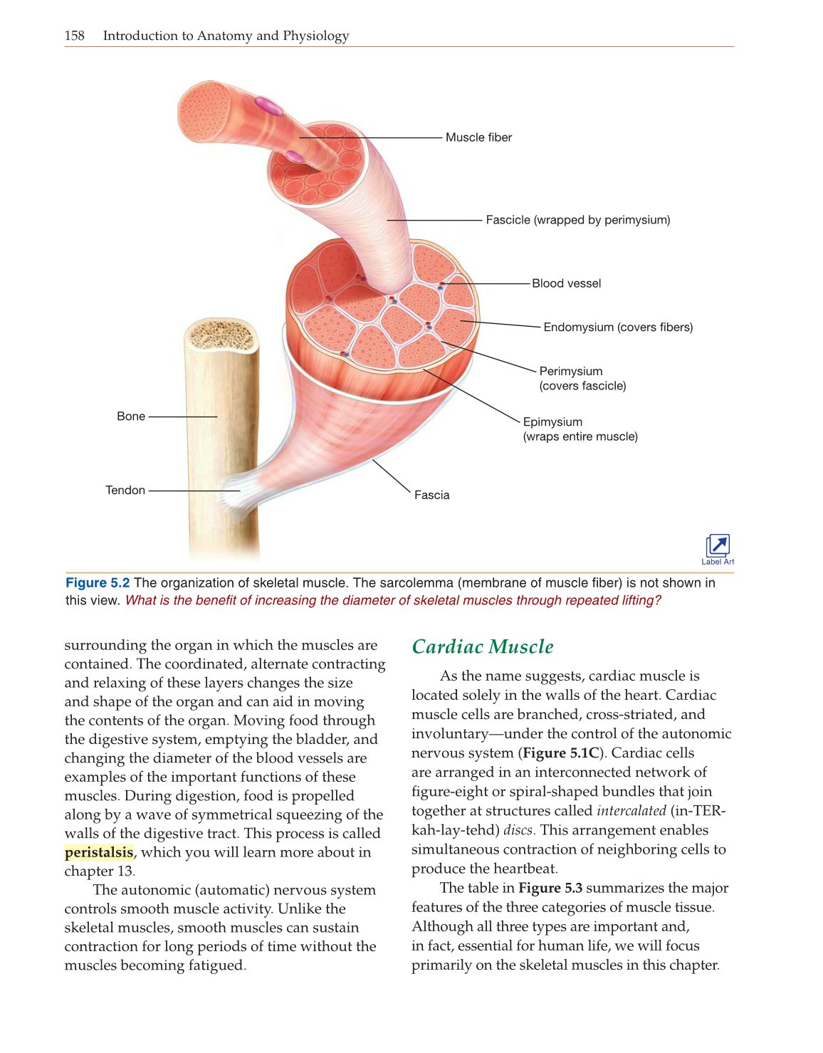
Figure 5.2 The organization of skeletal muscle. The sarcolemma (membrane of muscle fiber) is not shown in
this view. What is the benefit of increasing the diameter of skeletal muscles through repeated lifting?
Muscle fiber
Blood vessel
Fascia
Perimysium
(covers fascicle)
Epimysium
(wraps entire muscle)
Fascicle (wrapped by perimysium)
Endomysium (covers fibers)
Tendon
Bone
Label Art