164 Introduction to Anatomy and Physiology
the actin fi laments over the myosin fi laments,
resulting in a contraction of the sarcomere
(Figure 5.7).
What causes the actin fi laments to slide
over the myosin fi laments? Notice in Figure 5.7
that the myosin fi laments are encircled by small
protrusions called heads. When the sarcomere
is activated by an action potential, these heads
attach to receptor sites on the actin fi laments,
forming cross bridges. The cross bridges
contract, pulling the actin fi laments toward the
center of the sarcomere. During the process of
sarcomere contraction, these cross bridges attach,
pull, and release multiple times. The Ca++ ions
released with the arrival of the action potential
enable the attachments of the myosin heads to
the actin fi laments.
The neuromuscular system has the ability
to produce slow, gentle movements as well as
fast, forceful movements. This ability to produce
different kinds of movements and force variance
is accomplished by regulating the number and
frequency of action potentials. Only a small
number of action potentials are needed for
slow, gentle movements, while fast or forceful
movements require a large number of action
potentials, released rapidly.
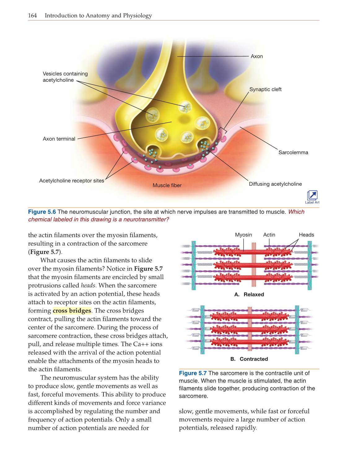
Figure 5.6 The neuromuscular junction, the site at which nerve impulses are transmitted to muscle. Which
chemical labeled in this drawing is a neurotransmitter?
Figure 5.7 The sarcomere is the contractile unit of
muscle. When the muscle is stimulated, the actin
filaments slide together, producing contraction of the
sarcomere.
Myosin Actin Heads
A. Relaxed
B. Contracted
Axon
Axon terminal
Vesicles containing
acetylcholine
Synaptic cleft
Sarcolemma
Muscle fiber
Acetylcholine receptor sites
Diffusing acetylcholine
Label Art